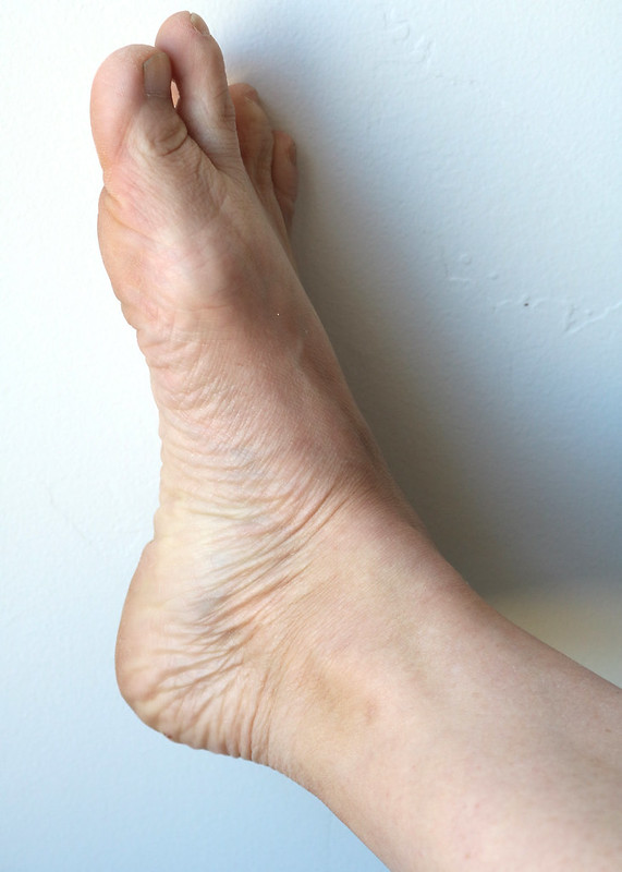
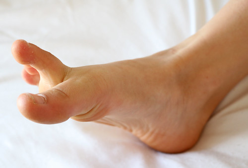
This video comprehensively explains the anatomical structure of the feet, with an emphasis on the bones. Bones and landmarks covered include Achilles tendon, calcaneus, phalanges, metatarsals, wrinkles, and veins.
- Watch the 57 min. version (Anatomy Lecture)
- Watch the Draw Along
- Watch the 24 second short
Other topics, such as how the stance of the feet affect the entire gesture of a figure, are also explored. Lecture by Art Prof Clara Lieu.
Video Walkthrough
- Feet can tell a story
- The feet “explain” the pose & weight of a figure
- The foot is more 3D than you think!
- Skip the foot muscles, rely on bones
- Most people draw the feet too small & too flat
- To learn proportions, draw complete figures. No missing body parts!
- The heel anchors the foot
- The heel shows weight distribution
- Look for planes on the bottom of the foot
- Group the little toes
- The big toe is a “loner”
- Notice how toes bend
- The big toe bends up, the little toes bend down
- Don’t trace toe nails, show how they affect the form
- Wrinkles & veins in the foot
- Follow the wrinkles in the foot
- Veins on the foot are prominent
Prof Lieu’s Tips

I think the biggest issue with feet I usually see when teaching is 1) drawing them too small, and 2) drawing them too flat.
Usually the reason why the feet are drawn too flat is people forget about the cluster of bones at the top of the foot, which are surprisingly substantial and dimensional.
I rarely tell people that the feet in their drawings are too small. Even when you think you are drawing them too large, they likely aren’t!

Bones mentioned
- Bone Groups: Talus, Cuboid, Cuneiform, Navicular
- Calcaneus
- Metatarsals
- Phalanges
- Medial malleolus
- Lateral malleolus
- Femur, Tibia, Fibula
Landmarks & Muscles mentioned
- Gastrocnemius
- Achilles Tendon
Artists mentioned
As a free educational source, Art Prof uses Amazon affiliate links (found in this page) to help pay the bills. This means, Art Prof earns from qualifying purchases.


